OptiBind™ Antibody Binder
Overview
WHY is Western blotting taking so long time?
WHY can’t we do antibody reactions more efficiently?
Here is the SOLUTION.
Now reduce antibody usage, reduce processing time!
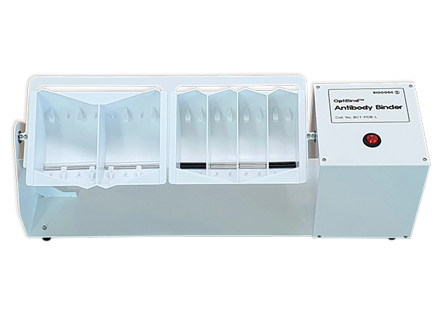
BCT-POB-L
Catalog Number
1 set
Unit Size
4℃ ~ RT
Storage
Features:
Developed faster Western Blotting results
Reduce the use of antibodies
Enhance Western Blotting signals
Features
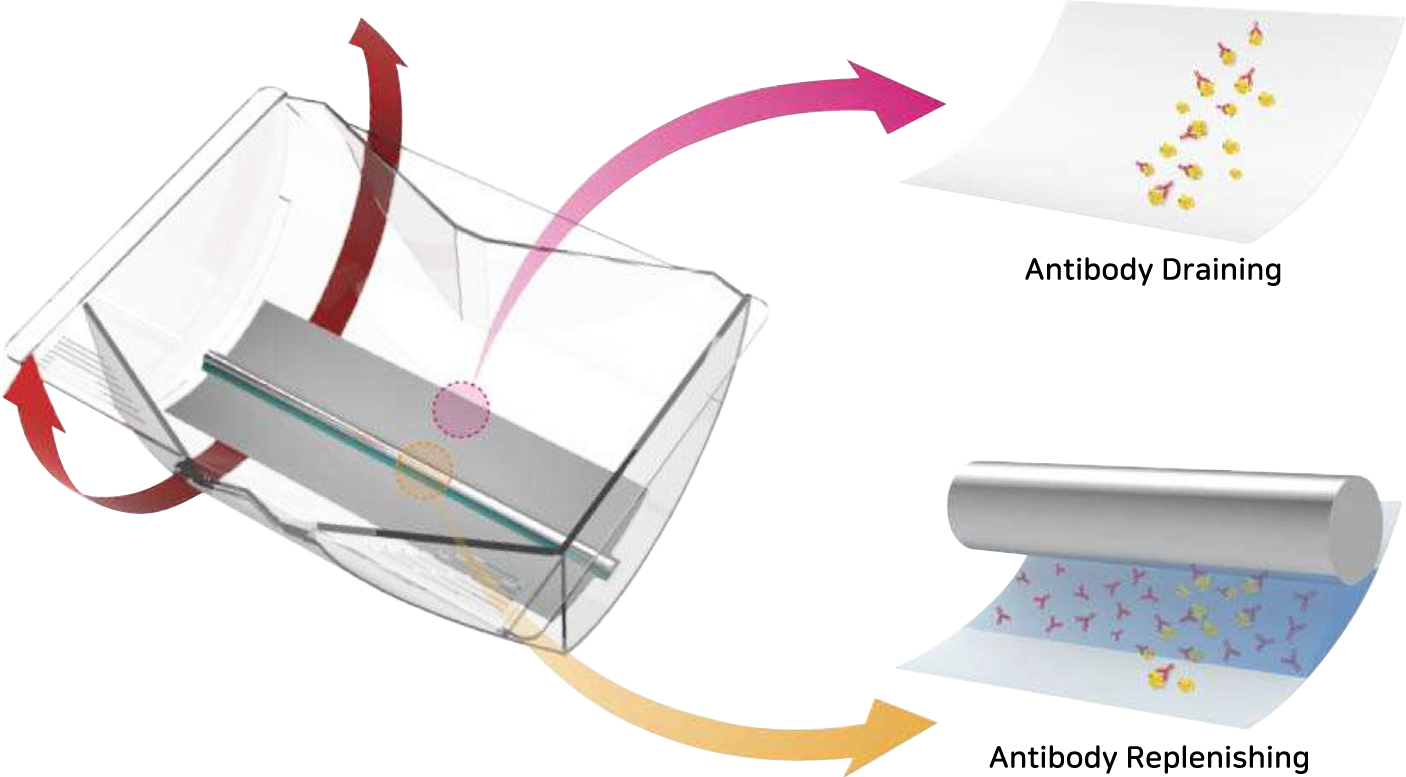
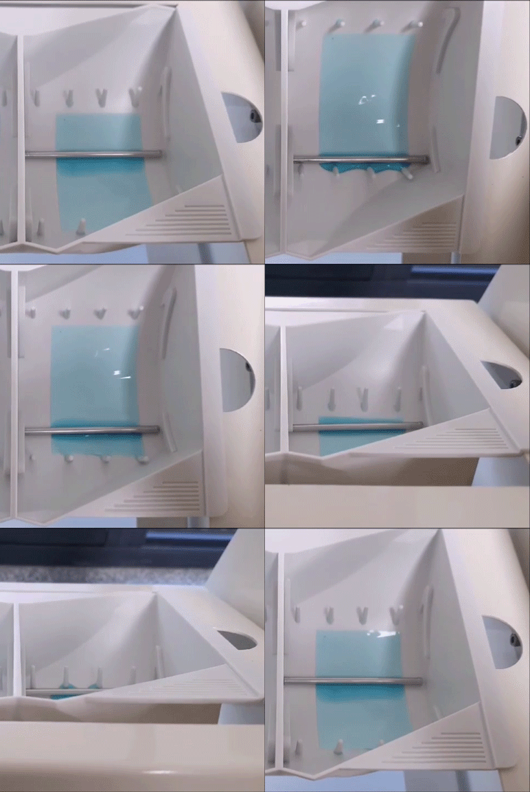
Antibody usage has decreased, how can the results be the same?
The Western blot is a simple and routine procedure, but each steps take hours or even days to complete. This is mainly due to mass transport limitations during the antibody incubation. When antibodies are introduced to the blotting membrane, they quickly attach to their targets–antigenic proteins– leaving a thin depletion layer along the surface. For Western blot, this makes a diffusion constraint for unbound antibody molecules to their antigens and hence the reaction takes hours to obtain enough signal. It has been recently proposed that this depletion layer can be easily disrupted by cyclic draining and replenishing (CDR) of the probe solution from the surface. A new open bucket-style chamber was devised for easier operation and the possibility of process automation. Rocking it back and forth achieved the CDR antibody incubation (R-CDR). The chamber was then equipped with a spreader-rod to facilitate the uniform movement of the antibody solution across the membrane surface. Hence, it was named spreader CDR (S-CDR). Compared to the batch incubation method, the S-CDR device produced significantly enhanced signals and developed faster results. There were several additional benefits of using the spreader-rod, which included uniform antibody binding across the membrane, reduced usage of antibodies, and the ability to recover results even from mishandled, creased membranes. The S-CDR device ensures better blots and can be easily implemented in existing western blot protocols.
Experimental Data

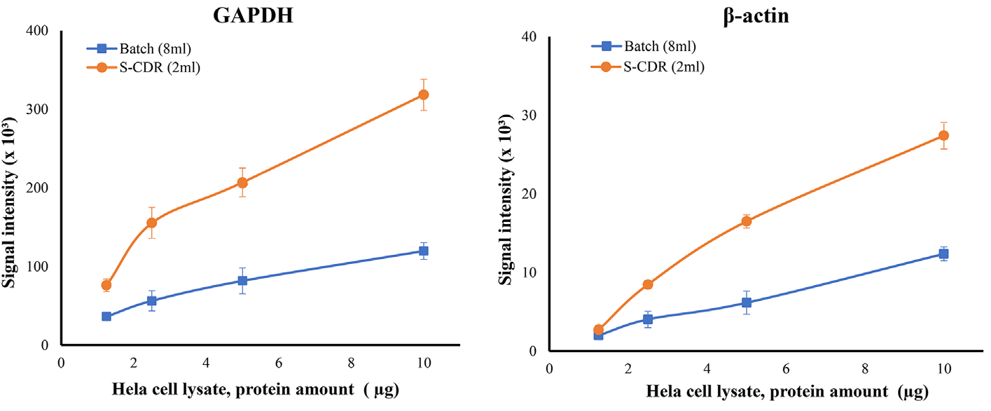
Differences in the results of Batch and S-CDR methods according to the sample concentration
Although the sample amount is small,
Although reducing the amount of antibodies used
The result is GOOD!
Sample: HeLa cell lysate (1.25 – 10 ug)
Membrane: NC membrane
Primary antibody: anti-β-actin, anti-GAPDH
Secondary antibody: IR Dye 680 LT goat anti-mouse IgG, IR Dye 800CW goat anti-rabbit IgG
Antibody incubation time: 45 min
Washing: TBS-T
Imaging: Odyssey Classic imaging system (LI-COR)

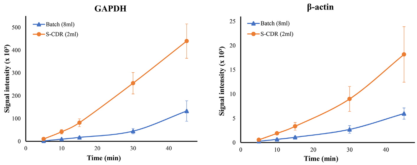
Differences in the results of Batch and S-CDR methods according to antibody incubation time
Reduce antibody reaction time
Sample: HeLa cell lysate
Membrane: NC membrane
Primary antibody: anti-β-actin, anti-GAPDH
Secondary antibody: IR Dye 680 LT goat anti-mouse IgG, IR Dye 800CW goat anti-rabbit IgG
Antibody incubation time: 5~45 min
Washing: TBS-T
Imaging: Odyssey Classic imaging system (LI-COR)
With Cover, there is no problem with long-term reaction.
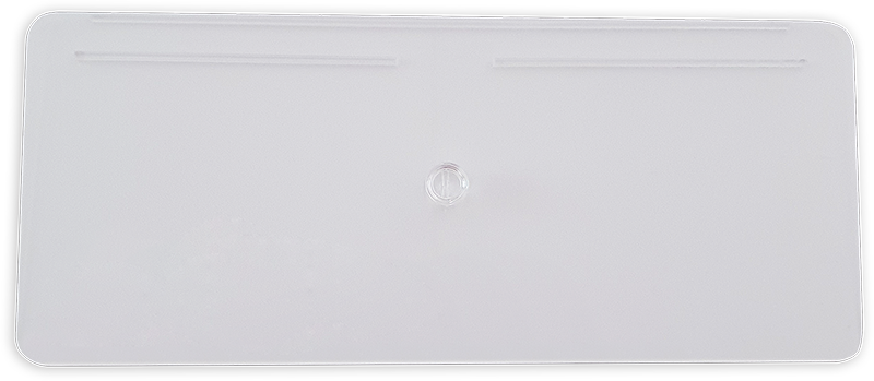
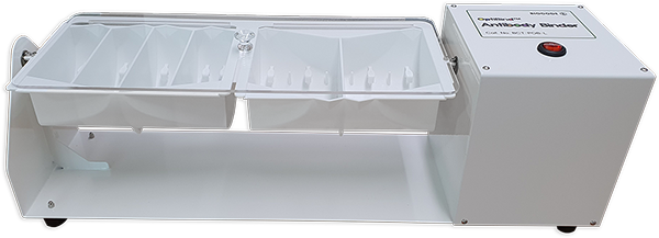
Cover (optional) can be used together to prevent the antibody solution from evaporating even in long-term reactions.
Cover is a transparent material, Even if you cover the device, you can see the membrane with the naked eye.
Specifications
| OptiBind™ Antibody Binder | |
|---|---|
| Angle | 45 ± 2° |
| Speed | 12 rpm |
| Basket size | Midi size: 85 × 150 mm/space Mini size: 85 × 75 mm/space Ultra mini size: 85 × 35 mm/space |
| Power | 220 V, 60 Hz |
| Dimensions | 560 (L) × 155 (D) × 183 (H) mm |
| Weight | 4.25 kg |
Ordering Information
| Code No. | Product | Unit |
|---|---|---|
| BCT-POB-L | OptiBind™ Antibody Binder | 1 set |
| BCT-SW-D | OptiBind™ Antibody Binding Basket, Midi-size | 1 ea |
| BCT-SW-N | OptiBind™ Antibody Binding Basket, Mini-size | 1 ea |
| BCT-SW-F | OptiBind™ Antibody Binding Basket, Ultra Mini-size | 1 ea |
| BCT-SW-C | OptiBind™ Antibody Binding Basket Cover | 1 ea |
